Seeing a problem can make a significant difference in your ability to understand it. It can be hard to identify a broken piece or misalignment if you cannot see the structures that are affected. Medical diagnostic imaging has made it possible to see your internal body parts, allowing experts to better understand conditions you may have. Fluoroscopy is one of these visual diagnostic aids.
Seeing Is Understanding
Fluoroscopy uses a continuous X-ray beam to show images of moving body parts—similar to a movie or recording of your internal structure’s functions. These images are projected onto a screen, allowing the doctor to see and analyze your body movements. When you have a painful biomechanical issues, seeing what your feet are doing when you use them can help our podiatrists identify the condition more accurately. This is particularly helpful for issues with fractures or dislocations.
The procedure can also help our doctors to identify and locate foreign objects, guide direct injections of medications into joints, give greater clarity for surgery, and check for circulatory issues. Foreign bodies and soft tissue structures coated in contrast dye allow the area being examined to appear bright white on the screen, so they stand out in the picture.
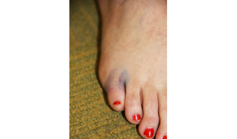
This can be helpful when treating and repairing chronic plantar fasciitis, Achilles tendinitis, neuromas, bunions, tailor’s bunions, and even hammertoes. The moving images allow the doctors to see the function—and damage—within the soft connective and nervous tissues. It also provides a closer look at the biomechanics that are affecting bony issues like bunions and bunionettes. That way, if surgery or other invasive actions are needed, our team is able to identify and locate the specific problem to treat it more accurately.
Risks Involved
Because fluoroscopy involves radiation, there are some risks. Extended periods of radiation or high-intensity beams are unhealthy for your body tissues. Fortunately, modern medicine allows lower levels of X-rays to be used while still getting a crisp, clear image. The procedure is now quite safe, limiting your odds for developing radiation side-effects later on. Our team also takes into account your medical history and previous exposure to X-rays, determining if you are healthy and able to absorb the beams without damage.
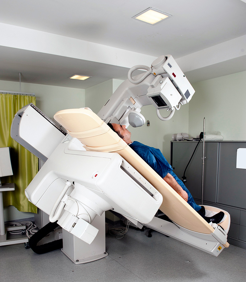
Performing the Procedure
If fluoroscopy is used to help determine your treatment, our team will discuss the risks and benefits with you. Once you decide to go ahead, we will prepare your feet for the X-rays. You will be positioned on a table and given directions about movement. We will inject you with contrast, so the necessary tissues appear in the images. The machine will be directed toward a specific area of your lower limbs and turned on. The doctor will study the images that result, and then move forward with any additional treatment or procedures. How long all of this takes will depend on your condition and unique needs, but we will not expose you to unnecessary radiation.
As medicine advances, we are able to provide more and more accurate information and remedies for our patients. Diagnostic imaging like fluoroscopy is valuable for helping us identify the best way to deal with chronic pain or other issues in your lower limbs. If you’d like to know more about this procedure, or would like any help with your lower limbs, contact Country Foot Care at either of our offices or use our REQUEST AN APPOINTMENT button at the top of this page.